Glossary of Genitourinary System Terms and Terminology
- Kidneys: The lima bean shaped bilateral urinary system organs that anatomically lie in the upper abdominal area in close proximity to the organs of the gastrointestinal system and just below the bilateral adrenal glands. These organs perform several roles in the body.
- The acid-base balance: The pH balance of the blood
- Erythropoietin: The substance that is needed for the production of red blood cells
- Calcitriol: The substance that is needed for the reabsorption of calcium, respectively.
- Renal cortex: The outer layer of the kidneys
- Renal medulla: The inside portion of the kidneys
- Filtration: The renal process that removes proteins and other cellular particles from the blood to create ultrafiltrate
- Reabsorption: The kidney process that entails the reentry of some particles and molecules from the ultrafiltrate back into the blood for future use
- Secretion: The renal process which entails the movement of wastes and other molecules into the urine from the blood after it is processed by the kidney
- Antidiuretic hormone: The hormone secreted by the pituitary gland that which controls the amount of water in the body and the blood and also decreases the amount of aldosterone in the body
- Vasopressin: An alternative name for antidiuretic hormone
- Aldosterone: The hormone that is secreted by the cortex of the adrenal gland, acts on the kidney to save sodium and water or to rid the body of sodium and water which affect the blood pressure
- Renin: The hormone that supports the cardiac system's blood pressure, electrolytes and circulating blood volume at the level of the kidneys.
- Angiotensin: A vasoconstrictor which increases the blood pressure and it also stimulates the release of aldosterone
- The Loop of Henle: A renal structure The Loop of Henle, as a part of the nephrons, manages the pressure differences that move fluids and molecules, such as electrolytes, across a membrane.
- Ureters: They connect the pelvis of each kidney to the bladder
- The bladder: The muscular organ that serves as the collection and retention vessel of the body.
- Micturition: The synonym for urination or voiding
- Urethra: The connection between the bladder and the external environment after the urine passes through the urinary sphincter muscle(s) and the urinary meatus, or opening.
- Urinary tract infection: A commonly occurring infection of the urinary tract
- Urolithiasis: A kidney stone or calculi
- Nephrolithiasis: Kidney stone or calculi
- Ureterolithiasis: A stone or calculus in the ureter
- Renal failure: A severe acute or chronic failure of the kidney to function adequately
- Renal cysts: Infections in the kidney
- Glomerulonephritis: Inflammation of the glomeruli of the kidneys
- Bladder tumors: Benign or malignant tumors in the bladder
- Cancers of the urinary tract: Cancers along the urinary tract
The Role of the Urinary System
The urinary system is also referred to as the renal system as well as the genitourinary system when structures and organs common to both the urinary system and the reproductive system are under consideration.
The urinary system plays several highly important roles in the body. For example, the urinary system:
- Rids wastes from the body
- Manages and controls the homeostasis of the body in terms of its pH or acid-base balance
- Manages and controls the homeostasis of the body in terms of its electrolytes and electrolyte balance
- Manages and controls the blood pressure
- Manages and controls the fluids and fluid balance in the body
- Filters the blood in the body
- Stores urine until voiding is prompted
- Enables the process of urination
The Parts of the Urinary System
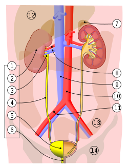
1. Human urinary system: 2. Kidney, 3. Renal pelvis, 4. Ureter, 5. Urinary bladder, 6. Urethra. (Left side with frontal section)
7. Adrenal gland
Vessels: 8. Renal artery and vein, 9. Inferior vena cava, 10. Abdominal aorta, 11. Common iliac artery and vein
With transparency: 12. Liver, 13. Large intestine, 14. Pelvis
The order of impurities being excreted from the kidneys: Kidneys → Ureters → Urinary Bladder → Urethra
The parts of the urinary system for both genders, as shown in the picture above, include the:
- Kidneys
- Ureters
- Bladder
- Urethra
All of these parts of the urinary system perform different roles in terms of the multiple roles of the urinary system, as described immediately above.
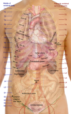
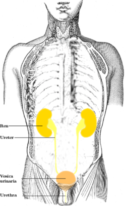
Surface projections of the organs of the trunk, showing kidneys at the level of T12 to L3.
The Kidneys
The kidneys, as shown in the picture above, are lima bean shaped bilateral urinary system organs that anatomically lie in the upper abdominal area in close proximity to the organs of the gastrointestinal system such as the stomach and liver and just below the bilateral adrenal glands which was discussed previously with the Endocrine System.
The kidneys are one of the most important organs of the body. Without adequate and normal kidney function a person will go into renal failure which can lead to death, have the need for renal, or kidney, dialysis or the need for kidney transplantation.
At least one functioning kidney is necessary to sustain life unless dialysis is done to perform the lost functions of the diseased or damaged kidney.
Physiologically and under normal conditions with healthy kidneys, these life sustaining urinary system organs filter the blood and:
- Remove wastes like urea and ammonia from the blood
- Manage and control the fluids and fluid balance in the body by holding or retaining water and releasing and removing water from the blood stream
- Manage and control the electrolyte balance of the blood
- Manage and control the acid-base or pH balance of the blood
- Serve with endocrine functions such as the production of erythropoietin and calcitriol which are needed for the production of red blood cells and the reabsorption of calcium, respectively.
The different sections of the kidney, as shown in the picture below. The two main layers of the kidney are the renal, which means kidney, medulla and the renal cortex. The renal medulla is the inside portion of the kidney; and the renal cortex is the outer layer of the kidney.
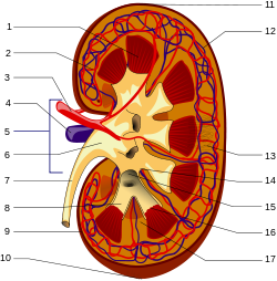
- Renal pyramid • 2. Interlobular artery • 3. Renal artery • 4. Renal vein 5. Renal hilum • 6. Renal pelvis • 7. Ureter • 8. Minor calyx • 9. Renal capsule • 10. Inferior renal capsule • 11. Superior renal capsule • 12. Interlobular vein • 13. Nephron • 14. Renal sinus • 15. Major calyx • 16. Renal papilla • 17. Renal column
The inside of the kidney, the renal medulla, holds the renal pyramids, as shown in the picture above. The kidneys houses millions of nephrons with are the primary functional cells of the kidney and the renal system.
Physiology of the Kidneys
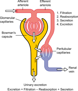
The physiological functioning of the kidneys in terms of its major functions relating to excretion and the fluid balance in the body, as shown in the pump like diagram above, occurs as the result of three functions, often in coordination with other mechanisms such as some endocrine glands' hormones.
These three functions are:
- Filtration
- Reabsorption
- Secretion
Filtration, simply stated, occurs when the body's circulating blood passes through the kidney and the filtration process removes proteins and other cellular particles from the blood to create ultrafiltrate, which eventually becomes urine after reabsorption and secretion occur.
Reabsorption occurs after filtration and reabsorption; reabsorption entails the reentry of some particles and molecules from the ultrafiltrate back into the blood for future use.
Secretion, simply stated, is the opposite of reabsorption. Secretion entails the movement of wastes and other molecules into the urine from the blood after it is processed by the kidney.
The process of urine production, in the correct sequential order is:
- The entry of circulating blood into the kidneys
- The filtration of the blood
- The production of ultrafiltrate
- Reabsorption from the blood
- Secretion into the blood
- Excretion of urine from the body
The role of the kidney in terms of cardiac and fluid balance entails its physiological prompting by endocrine hormones and enzymes including:
- Antidiuretic hormone
- Aldosterone which is a hormone
- Renin which is an enzyme
- Angiotensin II which is an enzyme
Antidiuretic hormone, as the name suggests prevents (anti) diuresis or urine production. Antidiuretic hormone, which is secreted by the pituitary gland, is also referred to as vasopressin which again, as the word suggests, vasopressin is a vascular (vaso) presser or squeezer (pressin). Antidiuretic hormone controls the amount of water in the body and the blood. Excessive water in the blood causes high blood pressure and fluid overload, and a low amount of water in the blood causes low blood pressure and dehydration. Antidiuretic hormone also decreases the amount of aldosterone in the body.
Aldosterone, which is secreted by the cortex of the adrenal gland, acts on the kidney to save sodium and water or to rid the body of sodium and water. When aldosterone reabsorbs sodium and fluid at the level of the kidneys, the blood pressure and blood volume increase; and conversely, when the circulating fluid and the blood pressure rise, aldosterone rids the body of sodium and water to decrease the blood pressure and to maintain the homeostasis of the body in terms of its blood pressure.
Renin, in concert with angiotensins, as shown the picture below, are the parts of the Renin-Angiotensin cycle which also supports the cardiac system's blood pressure, electrolytes and circulating blood volume at the level of the kidneys.
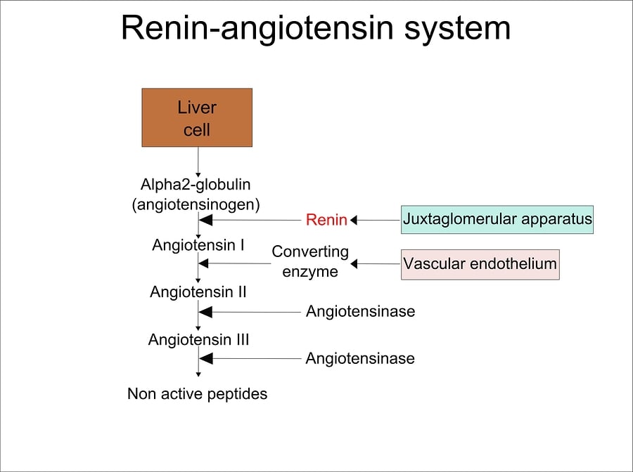
Scheme of the functioning of renin-angiotensin system.
The renin-angiotensin-aldosterone axis—that mediates extracellular volume (i.e., that of the blood plasma, lymph and interstitial fluid), and arterial vasoconstriction.
The enzyme renin, as stimulated or suppressed, is produced by the kidneys according to the blood pressure. Renin reduces the blood pressure when hypertension is present.
Angiotensin, another substance in the Renin-Angiotensin or the Renin-Angiotensin-Aldosterone subsystem, acts as a vasoconstrictor which increases the blood pressure and it also stimulates the release of aldosterone, as described above.
The Loop of Henle, as depicted in the diagram below, is another major anatomical and physiological part of the kidney and the renal functioning. The Loop of Henle, as a part of the nephrons, manages the pressure differences that move fluids and molecules, such as electrolytes, across a membrane.
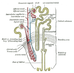
Scheme of renal tubule and its vascular supply. (Loop of Henle visible center-left.)
The Ureters

Ureter (Schematic View)
1. Human urinary system: 2. Kidney, 3. Renal pelvis, 4. Ureter, 5. Urinary bladder, 6. Urethra. (Left side with frontal section), 7. Adrenal gland
Vessels:
8. Renal artery and vein, 9. Inferior vena cava, 10. Abdominal aorta, 11. Common iliac artery and vein
With transparency:
12. Liver, 13. Large intestine, 14. Pelvis
The bilateral ureters connect the pelvis of each kidney to the bladder, as shown in the picture above. The ureters consist of smooth involuntary muscle which serves as the conduit of urine from the kidneys into the bladder. Unlike the kidney, the ureters do NOT play any physiological role in the functioning of other bodily systems like the cardiovascular system.
The Bladder
The bladder, as shown in the picture above, is the muscular organ that serves as the collection and retention vessel of the body. The bladder temporarily holds and retains urine prior to micturition which is a synonym for urination or voiding. Urine fills the bladder and the bladder stretches and increases in size as it receives urine from the ureters. The collected urine leaves the bladder through the urethra to the external environment with urination as the bladder contracts and decreases in terms of its size. This muscular organ can, under normal circumstances, comfortably hold about 500 to 800 milliliters (mLs) of urine.
On a daily basis, and under normal circumstances, humans void from 800 to 2,000 (mLs) of urine. Urinary output less than 800 mLs per day and less than 30 mLs per hour are considered oliguria; urinary output in excess of 2,000 mLs per day is considered polyuria; and the absence of all urine production is referred to as anuria.
The Urethra
The urethra differs among males and females as shown in the pictures below:
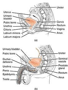
The urethra transports urine from the bladder to the outside of the body. This image shows (a) a female urethra and (b) a male urethra.
The urethra is the connection between the bladder and the external environment after the urine passes through the urinary sphincter muscle(s) and the urinary meatus, or opening.
Because the urethra, among males, transports both urine and sperm during ejaculation, the male gender has two urethral sphincters to control these two different and separate functions. Because females transport only urine through the urethra, the female gender has only one urethral sphincter to control the flow of urine only.
Disorders Affecting the Urinary System
- Urinary tract infection
- Urolithiasis
- Nephrolithiasis
- Ureterolithiasis
- Renal failure
- Renal cysts
- Glomerulonephritis
- Bladder tumors
- Cancers of the urinary tract
RELATED TEAS ANATOMY & PHYSIOLOGY CONTENT:
- General Anatomy and Physiology of a Human
- Respiratory System
- Cardiac System
- Circulatory System
- Digestive or Gastrointestinal System
- Nervous System
- Musculoskeletal System – Skeletal
- Musculoskeletal System – Muscular
- Reproductive System
- Integumentary System
- Endocrine System
- Genitourinary System (Currently here)
- Immune System
- Hematological System
